Bahjat Ghazzal

and 1 more

Atypical Atrial Flutter Ablation: The Clinical Impact of High-Density MappingBahjat Z. Ghazzal MD1, Marwan M. Refaat, MD21 Division of Cardiology, Department of Internal Medicine, University of Massachusetts Chan Medical School , Worcester, Massachusetts, USA2 Division of Cardiology, Department of Internal Medicine, American University of Beirut Medical Center, Beirut, LebanonRunning Title: Atypical Atrial Flutter Ablation: The Clinical Impact of High-Density MappingWords: 700 (excluding the title page and references)Keywords: cardiac arrhythmias, heart diseases, cardiovascular diseases, catheter ablation, atrial flutter, high density mappingFunding: NoneDisclosures: NoneCorresponding Author:Marwan M. Refaat, MD, FACC, FAHA, FHRS, FASE, FESC, FACP, FRCPTenured Professor of MedicineTenured Professor of Biochemistry and Molecular GeneticsMember, Division of Cardiology/ Section of Cardiac ElectrophysiologyDirector, Cardiovascular Fellowship Program American University of Beirut Faculty of Medicine and Medical CenterPO Box 11-0236, Riad El-Solh 1107 2020- Beirut, LebanonUS Address: 3 Dag Hammarskjold Plaza, 8th Floor, New York, NY 10017, USAOffice: +961-1-350000/+961-1-374374 Extension 5353 or Extension 5366 (Direct)Atypical atrial flutter (AAFL) is an arrhythmia that is distinct from typical atrial flutter (AFL) due to its non-isthmus-dependent reentrant circuit, and often arises in patients with a history of cardiac surgery or ablation, where the resulting iatrogenic scar provides the basis for re-entry circuits.1 Catheter ablation for AAFL is an effective treatment option, associated with lower rates of thromboembolic events, transfusions, and shorter hospital stays compared to typical AFL.2 However, the complexity of AAFL circuits requires precise mapping, and traditional techniques often lack the resolution to accurately delineate these pathways. High-density (HD) mapping technology has emerged as a significant advancement in this regard, offering detailed electroanatomic maps that enhance procedural success.3 In this issue of the Journal of Cardiovascular Electrophysiology, the study by Sink et al. investigates the impact of HD mapping on AAFL ablation outcomes and healthcare utilization, providing valuable insights into its clinical benefits.4In this retrospective analysis, Sink et al. examined 108 patients who underwent AAFL ablation at a single academic center from 2005 to 2022. The cohort was divided into two groups: those who received HD mapping with a 16-electrode HD Grid catheter and Precision mapping system, and those who underwent non-HD mapping using traditional spiral catheters and the Velocity system. Primary outcomes included procedural success, defined as non-inducibility of the arrhythmia after ablation, and AFL recurrence. Secondary outcomes included emergency department (ED) visits, hospitalizations, and overall healthcare utilization within one-year post-procedure. The study revealed that HD mapping significantly improved both the delineation of AAFL circuits (92.5% vs. 76%; p=0.014) and procedural success rates (91% vs. 71%; p=0.006). Additionally, patients in the HD mapping group experienced significantly fewer ED visits (aIRR 0.32; p=0.007) and hospitalizations (aIRR 0.32; p=0.004) for AF/AFL/HF within the first year. While there was a trend towards lower AFL recurrence in the HD mapping group (aHR 0.60; p=0.13), this difference did not reach statistical significance. This study highlights the significant advantages of HD mapping in AAFL ablation. The enhanced resolution provided by HD mapping allows for more precise identification and targeting of arrhythmogenic substrates, leading to improved clinical outcomes.Though this study was well-conducted, minor limitations must be noted. The study’s retrospective design and single-center setting introduce potential biases and limit the generalizability of the results. The non-randomized nature of the study also raises the possibility of selection bias. Furthermore, the higher use of contact force-sensing catheters in the HD mapping group may have contributed to the observed differences in outcomes. A larger sample size would have likely allowed the study to achieve statistical significance when comparing recurrent AAFL rates between groups. Despite these limitations however, this study provides strong support for the clinical utility of HD mapping in AAFL ablation. Future multi-center, randomized trials should validate these findings and examine factors not studied here, such as procedure fluoroscopy time and complication rates between groups. Long-term benefits should also be further investigated, as some prior studies for example have shown no significant decrease in anti-arrhythmic drug use at 1-year follow-up5 and significantly decreased rates of sinus rhythm maintenance for repeat ablations compared with single ablations6. Research on HD mapping in patients with specific comorbidities could also provide insights into its broader applicability. For instance, one study showed a greater chance of acute procedural failure in patients with a history of surgical correction for congenital heart disease.7 Though HD-mapping-guided ablation demonstrates higher acute procedural success, several studies have shown long-term recurrence rates remain significant.8, 9 Hence, further research is needed to identify strategies to reduce these rates. In this context, the development of new mapping tools, such as the Octaray TM system (Biosense Webster Inc., Irvine, CA, USA)10 and the Ensite TM Omnipolar Technology (OT) (Abbott, Chicago, IL, USA)11, may offer improved outcomes. Further research is also needed to assess the clinical outcomes, healthcare utilization and biomarker response for ablation of AAFL with HD mapping in the setting of heart failure with reduced ejection fraction.12 To the best of our knowledge, this is the first such study to demonstrate that ablation with HD mapping for AAFL results in reduced re-hospitalization and ED visits. Future research should attempt to expand on these findings to evaluate potential cost-benefit and impact on patient quality of life.References:1. Cherian Tharian S, Supple G, Smietana J, Santangeli P, Nazarian S, Lin D, Hyman Matthew C, Walsh K, Marchlinski F and Arkles J. Idiopathic Atypical Atrial Flutter Is Associated With a Distinct Atriopathy.JACC: Clinical Electrophysiology . 2021;7:1193-1195.2. Ko Ko NL, Sriramoju A, Khetarpal BK and Srivathsan K. Atypical atrial flutter: review of mechanisms, advances in mapping and ablation outcomes.Curr Opin Cardiol . 2022;37:36-45.3. Raymond-Paquin A, Pillai A, Myadam R, Mankad P, Lovejoy S, Koneru JN and Ellenbogen KA. Atypical atrial flutter catheter ablation in the era of high-density mapping.J Interv Card Electrophysiol . 2023;66:1807-1815.4. Sink JC, Kasen; Uppalapati, Lakshmi; Lancki, Nicola; Peigh, Graham; Lohrmann, Graham; Elsayed, Mahmoud; Carneiro, Herman; Baman, Jayson; Pfenniger, Anna; Patil, Kaustubha D.; Verma, Nishant; Arora, Rishi; Kim, Susan S.; Chicos, Alexandru B.; Lin, Albert C.; Knight, Bradley P.; Passman, Rod S. Association Between High-Density Mapping of Atypical Atrial Flutter, Clinical Outcomes and Healthcare Utilization. Journal of Cardiovascular Electrophysiology . 2024.5. Balt JC, Klaver MN, Mahmoodi BK, van Dijk VF, Wijffels MCEF and Boersma LVA. High-density versus low-density mapping in ablation of atypical atrial flutter.Journal of Interventional Cardiac Electrophysiology . 2021;62:587-599.6. Marazzato J, Cappabianca G, Angeli F, Crippa M, Golino M, Ferrarese S, Beghi C and De Ponti R. Catheter ablation of atrial tachycardias after mitral valve surgery: a systematic review and meta‐analysis. Journal of Cardiovascular Electrophysiology . 2020;31:2632-2641.7. Delacretaz E, Ganz Leonard I, Soejima K, Friedman Peter L, Walsh Edward P, Triedman John K, Sloss Laurence J, Landzberg Michael J and Stevenson William G. Multiple atrial macro–re-entry circuits in adults with repaired congenital heart disease: entrainment mapping combined with three-dimensional electroanatomic mapping.Journal of the American College of Cardiology . 2001;37:1665-1676.8. Lozano-Granero C, Moreno J, Sanchez-Perez I, Matia-Frances R, Hernandez-Madrid A, Zamorano JL and Franco E. Results of atypical flutter ablation in the era of high density electroanatomical mapping (the RAFAELA study). European Heart Journal . 2023;44.9. Anter E, McElderry TH, Contreras-Valdes FM, Li J, Tung P, Leshem E, Haffajee CI, Nakagawa H and Josephson ME. Evaluation of a novel high-resolution mapping technology for ablation of recurrent scar-related atrial tachycardias. Heart Rhythm . 2016;13:2048-2055.10. Sroubek J, Rottmann M, Barkagan M, Leshem E, Shapira‐Daniels A, Brem E, Fuentes‐Ortega C, Malinaric J, Basu S and Bar‐Tal M. A novel octaray multielectrode catheter for high‐resolution atrial mapping: electrogram characterization and utility for mapping ablation gaps. Journal of Cardiovascular Electrophysiology . 2019;30:749-757.11. Rillo M, Palamà Z, Punzi R, Vitanza S, Aloisio A, Polini S, Tucci A, Pollastrelli A, Zonno F, Anastasia A, Giannattasio CF and My L. A new interpretation of nonpulmonary vein substrates of the left atrium in patients with atrial fibrillation. Journal of Arrhythmia . 2021;37:338-347.12. Pascual-Figal D, Wachter R, Senni M, Bao W, Noè A, Schwende H, Butylin D, Prescott MF; TRANSITION Investigators. NT-proBNP Response to Sacubitril/Valsartan in Hospitalized Heart Failure Patients With Reduced Ejection Fraction: TRANSITION Study. JACC Heart Fail Oct 2020;8(10):822-833.

Bahjat Ghazzal

and 1 more

not-yet-known not-yet-known not-yet-known unknown The Efficiency of using KardiaMobile 6L in the Cardiac Electrophysiology Clinic Bahjat Z. Ghazzal MD1, Marwan M. Refaat, MD2 1 Division of Cardiology, Department of Internal Medicine, University of Massachusetts Chan Medical School , Worcester, Massachusetts, USA 2 Division of Cardiology, Department of Internal Medicine, American University of Beirut Medical Center, Beirut, Lebanon Running Title: The Efficiency of KardiaMobile 6L in Clinic Words: 719 (excluding the title page and references) Keywords: Electrocardiogram, cardiac arrhythmias, cardiology, cardiovascular diseases, Utilization Time, Efficiency Funding: None Disclosures: None Corresponding Author: Marwan M. Refaat, MD, FACC, FAHA, FHRS, FASE, FESC, FACP, FRCP Tenured Professor of Medicine Tenured Professor of Biochemistry and Molecular Genetics Member, Division of Cardiology/ Section of Cardiac Electrophysiology Director, Cardiovascular Fellowship ProgramAmerican University of Beirut Faculty of Medicine and Medical Center PO Box 11-0236, Riad El-Solh 1107 2020- Beirut, Lebanon US Address: 3 Dag Hammarskjold Plaza, 8th Floor, New York, NY 10017, USA Office: +961-1-350000/+961-1-374374 Extension 5353 or Extension 5366 (Direct) Electrocardiography (ECG) is an essential diagnostic tool in cardiology, allowing for the detection and management of various cardiac conditions. Traditional 12-lead ECGs, while comprehensive, can be time-consuming and may impact clinic efficiency. This may be particularly important in outpatient cardiology clinics, where time can often be a scarce resource. A survey distributed at the 2022 ESC congress found that while most cardiologists believe that consultations should last 30 to 45 minutes, they often have only 20 minutes or less.1 Although the effects of time pressure have not been extensively studied in cardiology and cardiac electrophysiology outpatient clinics, research in primary care has linked it to increased physician stress, burnout, intent to leave the practice2 in addition to lower diagnosis rates and higher follow-up care rates3. Research has also shown that increasing system efficiency and improving patient cycle time in primary care clinics can improve patient experience and access as well as staff satisfaction.4 Thus, the introduction of KardiaMobile 6-lead ECG (Figure 1), a more portable and user-friendly device, could present an opportunity to streamline ECG collection and potentially improve clinic time utilization. The study by Gaddam et al. explores this potential by comparing room utilization times between KardiaMobile 6-lead ECG and the standard 12-lead ECG in a cardiology clinic setting. In their study, Gaddam et al. conducted a single-center, non-randomized trial involving 100 patients aged 18 to 89, excluding those with resting tremor. The participants were divided into two groups: one underwent ECG collection using KardiaMobile 6-lead ECG, and the other using the traditional 12-lead ECG. The primary outcome measured was room utilization time, with secondary outcomes including the need for additional 12-lead ECGs. The results demonstrated a significant reduction in room utilization time with KardiaMobile 6-lead ECG (7.27 minutes) compared to the 12-lead ECG (10.33 minutes, p < 0.001). Only 16% of visits in the KardiaMobile 6-lead ECG group required an additional 12-lead ECG, indicating that KardiaMobile 6-lead ECG is sufficient for most clinical needs. The primary benefit of 6-lead ECGs lies in their portability and ease of use, making them suitable for continuous monitoring and rapid assessment in both clinical and non-clinical settings. Although research on this subject is still limited, a recent prospective study of 1,015 participants found that the KardiaMobile 6-lead ECG demonstrates a high level of agreement with 12-lead ECGs for certain parameters like PR interval, QRS duration, and cardiac axis, but may be less effective for detecting conditions that require detailed precordial lead information, such as left ventricular hypertrophy or specific ischemic changes.5 Other studies have also corroborated acceptable agreement in certain ECG parameters between 6-lead and 12-lead ECG’s, however, highlighting the 12-lead ECG’s advantage in certain situations where broader and more detailed data collection is required.6,7 While this study was well-conducted, minor limitations exist. The study follows a non-randomized design, which may potentially introduce selection bias and the team members were non-blinded, which may introduce observer bias. Additionally, the relatively small sample size and single-center nature of the study may limit generalizability. It must also be noted that nearly half of the patients in each group (KardiaMobile 6-lead ECG vs. 12-lead ECG) visited the clinic for atrial fibrillation and/or atrial flutter follow-up, conditions that can be detected with just a 1-lead ECG. Finally, while the study shows a statistically significant reduction in average room utilization time by 3.07 minutes, it does not assess whether this reduction translated to meaningful improvements in tangible clinic efficiency outcomes. Despite these limitations, the study provides valuable insights into the potential benefits of integrating KardiaMobile 6-lead ECG into clinical practice. It could lead to improved clinical workflows, allowing clinicians to see more patients without compromising the quality of care. This device may also be particularly beneficial in remote or resource-limited settings where traditional 12-lead ECGs are impractical. Future research should focus on larger, randomized trials to validate these findings and explore the long-term benefits and potential limitations of KardiaMobile 6-lead ECG in diverse clinical environments. Furthermore, investigating the use of KardiaMobile 6-lead ECG in specific patient populations, such as those with complex arrhythmias or comorbidities associated with arrhythmias such as cardiomyopathies and heart failure, could provide additional insights into its clinical utility.8,9 The development of guidelines and protocols for integrating KardiaMobile 6-lead ECG into routine practice will be essential to maximize its benefits and ensure patient safety. Legend Figure 1: Kardia 6L Electrocardiogram (Mountain View, CA) References: https://doi.org/10.1016/j.jelectrocard.2021.03.008 1. Sala O, Moscatelli S. Time matters. Global assessment of quality, duration and mismatch between real practice working conditions and physician needs performing outpatients cardiological consultations. European Heart Journal . 2022;43. doi: 10.1093/eurheartj/ehac544.28352. Prasad K, Poplau S, Brown R, Yale S, Grossman E, Varkey AB, Williams E, Neprash H, Linzer M, for the Healthy Work Place I. Time Pressure During Primary Care Office Visits: a Prospective Evaluation of Data from the Healthy Work Place Study. Journal of General Internal Medicine . 2020;35:465-472. doi: 10.1007/s11606-019-05343-63. Freedman S, Golberstein E, Huang TY, Satin DJ, Smith LB. Docs with their eyes on the clock? The effect of time pressures on primary care productivity. J Health Econ . 2021;77:102442. doi: 10.1016/j.jhealeco.2021.1024424. Robinson J, Porter M, Montalvo Y, Peden CJ. Losing the wait: improving patient cycle time in primary care. BMJ Open Quality . 2020;9:e000910. doi: 10.1136/bmjoq-2019-0009105. Azram M, Ahmed N, Leese L, Brigham M, Bowes R, Wheatcroft SB, Ngantcha M, Stegemann B, Crowther G, Tayebjee MH. Clinical validation and evaluation of a novel six-lead handheld electrocardiogram recorder compared to the 12-lead electrocardiogram in unselected cardiology patients (EVALECG Cardio). European Heart Journal - Digital Health . 2021;2:643-648. doi: 10.1093/ehjdh/ztab0836. Madias JE. A Comparison of 2-Lead, 6-Lead, and 12-Lead ECGs in Patients With Changing Edematous States: Implications for the Employment of Quantitative Electrocardiography in Research and Clinical Applications. CHEST . 2003;124:2057-2063. doi: 10.1378/chest.124.6.20577. Orchard JJ, Orchard JW, Raju H, La Gerche A, Puranik R, Semsarian C. Comparison between a 6‑lead smartphone ECG and 12‑lead ECG in athletes. Journal of Electrocardiology . 2021;66:95-97. doi: 8. El Moheb M, Nicolas J, Khamis AM, Iskandarani G, Akl EA, Refaat M. Implantable Cardiac Defibrillators for patients with non-ischaemic cardiomyopathy. Cochrane Database Syst Rev Dec 2018; 12: CD012738 9. Pascual-Figal D, Wachter R, Senni M, Bao W, Noè A, Schwende H, Butylin D, Prescott MF; TRANSITION Investigators. NT-proBNP Response to Sacubitril/Valsartan in Hospitalized Heart Failure Patients With Reduced Ejection Fraction: TRANSITION Study. JACC Heart Fail. Aug 2020; S2213-1779(20)30336-X.

Alicia Khazzeka

and 9 more

Introduction: The aim of this study was to describe our experience and outcome of ablation therapy of arrhythmias in children at a tertiary care center. Methods: Data was collected retrospectively from the hospital medical records. All children presenting to AUBMC between 2000 and 2020 who underwent cardiac ablation were included. The data collected included type of arrhythmia, ablation technique, age and weight at ablation, procedure complications, medications used, and outcome assessment. Results: We had 67 patients who underwent cardiac ablation. Of those, 60% were males with a mean age of 15 years. Structural heart disease was present in 6% of patients. Wolff-Parkinson-White syndrome (WPW) was most prevalent at 31%, followed by atrioventricular nodal reentrant tachycardia (AVNRT) at 24%, atrioventricular reentrant tachycardia (AVRT) at 19%, ventricular tachycardia (VT) at 10%, atrial fibrillation (AF) at 2%, and atrial tachycardia (AT) at 1%. The remaining 13% of patients presented with less common types of arrhythmias, including narrow complex tachycardia, retrograde dual atrioventricular nodal reentry, premature ventricular contractions (PVC), and orthodromic reciprocating tachycardia. Antiarrhythmic medications were started prior to the procedure in 59% of our population. Medication regimens post-ablation included beta blockers (68%), type 1c antiarrhythmics (25%), calcium channel blockers (3%), ivabradine (2%), and amiodarone (2%). The completed procedures showed a success rate of 93%. Conclusion: Ablation of arrhythmias in children is an effective procedure in the treatment of childhood arrhythmias. More studies are needed on cardiac ablation in children with structural heart disease in the Middle East region.

Diane Rizkallah

and 1 more

Safety and Effect on Length of Stay of Intravenous Sotalol Initiation for Arrhythmia ManagementDiane H. Rizkallah, BS; Marwan M. Refaat, MDDivision of Cardiology, Department of Internal Medicine, American University of Beirut Medical Center, Beirut, LebanonRunning Title: Safety and Effect on LOS of IV SotalolWords: 598 (excluding the title page and references)Keywords: Heart Diseases, Cardiovascular Diseases, Cardiac Arrhythmias, Safety, Length of StayFunding: NoneDisclosures: NoneCorresponding Author:Marwan M. Refaat, MD, FACC, FAHA, FHRS, FRCPTenured Professor of MedicineTenured Professor of Biochemistry and Molecular GeneticsVan Dyck Medical Educator and Director of the Cardiovascular Fellowship ProgramDepartment of Internal Medicine, Cardiovascular Medicine/Cardiac ElectrophysiologyAmerican University of Beirut Faculty of Medicine and Medical CenterPO Box 11-0236, Riad El-Solh 1107 2020- Beirut, LebanonUS Address: 3 Dag Hammarskjold Plaza, 8th Floor, New York, NY 10017, USAOffice: +961-1-350000/+961-1-374374 Extension 5353 or Extension 5366 (Direct)Email: [email protected] is a class III antiarrhythmic drug with beta-adrenergic blocking activity, used to manage both supraventricular and ventricular arrhythmias. It is available in both oral and intravenous formulations[1]. The FDA approved intravenous Sotalol in March 2020. Sotalol is known to cause QTc prolongation with serum sotalol concentration linearly correlating with QTc length regardless of the route of administration [2,3], with women being at higher risk than men[4]. QTc interval prolongation is one of many parameters that is associated with cardiovascular mortality [5]. QTc prolongation may lead to polymorphic ventricular tachycardia/Torsade de Pointes which is a potentially lethal condition that is acquired from medications or due to an underlying channelopathy predisposing to sudden cardiac arrest[6]. Additional adverse effects of sotalol may include hypotension, bradycardia and AV block[7,8]. Nevertheless, since its approval, IV sotalol has been successfully and safely used in both adult and pediatric patient populations for the management of arrhythmias in acute and chronic settings [9,10,11,12]. Initiation of sotalol therapy with PO loading requires 5 successive oral doses over a 3-day hospital stay for monitoring, at an estimated cost of $2931.55 per day [13]. In 2020, a protocol for IV loading of sotalol was developed using data modeling. This protocol was hypothesized to allow a significant reduction in the length of hospitalization, and thus in the incurred costs[14]. However, there has not been any large-scale implementation of this protocol, nor any comparison of its safety profile and efficacy to that of the traditional oral loading protocol.This study by Liu et al. is a nonrandomized clinical trial in which 29 patients underwent IV sotalol loading. They were compared by chart review to 20 patients who underwent PO sotalol loading in the same timeframe. The indication for sotalol initiation in both cases was for primary atrial or ventricular arrhythmias. The study’s main aim was to assess the safety profile of IV sotalol loading while comparing the length of hospitalization to that required for PO sotalol loading. The same exclusion and inclusion criteria were applied to both groups. Notably, patients with significantly depressed LVEF and creatinine clearance were excluded. The study revealed that safety outcomes were similar in both groups but that IV sotalol loading led to significantly shorter hospital stays. It also found that QT or QTc in patients receiving IV sotalol was similar at the conclusion of the one-hour infusion to that at discharge.These findings support the use of IV loading for sotalol initiation, as they suggest it requires shorter hospital stays than PO loading with similar safety profiles. As shorter hospital stays translate into lower patient days, lower costs, and these results suggest IV sotalol loading is more cost-efficient than its oral counterpart. They also suggest that the maximal increase in QT or QTc length following sotalol initiation is attained by the end of IV loading, thus indicating that patients may be discharged within less than 24 hours of drug initiation.While this study offered valuable insight, its design had significant limitations. Firstly, this was not a randomized clinical trial. A comparison of baseline characteristics between the two populations studied revealed a significantly higher proportion of females in the oral group, which may have inherently skewed outcomes related to QT and QTc length.Secondly, the sample size was small, with no long-term follow-up. Lastly, patients with significantly depressed GFR or LVEF, particularly prone to developing adverse effects with sotalol use, were excluded from the study. Randomized clinical trials examining the short-term and long-term safety of IV sotalol loading and the optimal length of hospitalization are needed, and such efforts are already underway.References:Batul, S. A., & Gopinathannair, R. (2017). Intravenous Sotalol - Reintroducing a Forgotten Agent to the Electrophysiology Therapeutic Arsenal. Journal of atrial fibrillation , 9 (5), 1499. https://doi.org/10.4022/jafib.1499Somberg, J. C., Preston, R. A., Ranade, V., & Molnar, J. (2010). QT prolongation and serum sotalol concentration are highly correlated following intravenous and oral sotalol. Cardiology , 116 (3), 219–225. https://doi.org/10.1159/000316050Barbey, J. T., Sale, M. E., Woosley, R. L., Shi, J., Melikian, A. P., & Hinderling, P. H. (1999). Pharmacokinetic, pharmacodynamic, and safety evaluation of an accelerated dose titration regimen of sotalol in healthy middle-aged subjects. Clinical pharmacology and therapeutics , 66 (1), 91–99. https://doi.org/10.1016/S0009-9236(99)70058-5Somberg, J. C., Preston, R. A., Ranade, V., Cvetanovic, I., & Molnar, J. (2012). Gender differences in cardiac repolarization following intravenous sotalol administration. Journal of cardiovascular pharmacology and therapeutics , 17 (1), 86–92. https://doi.org/10.1177/1074248411406505Al-Kindi SG, Refaat M, Jayyousi A, Asaad N, Al Suwaidi J, Abi Khalil C. Red Cell Distribution Width is Associated with All-Cause and Cardiovascular Mortality in Patients with Diabetes. Biomed Res Int 2017; 2017: 5843702Refaat MM, Hotait M, Tseng ZH: Utility of the Exercise Electrocardiogram Testing in Sudden Cardiac Death Risk Stratification.Ann Noninvasive Electrocardiol 2014; 19(4): 311-318.Marill, K. A., & Runge, T. (2001). Meta-analysis of the Risk of Torsades de Pointes in patients treated with intravenous racemic sotalol. Academic emergency medicine,  8 (2), 117–124. https://doi.org/10.1111/j.1553-2712.2001.tb01275.xMacNeil, D. J., Davies, R. O., & Deitchman, D. (1993). Clinical safety profile of sotalol in the treatment of arrhythmias. The American journal of cardiology , 72 (4), 44A–50A. https://doi.org/10.1016/0002-9149(93)90024-7Malloy-Walton, L. E., Von Bergen, N. H., Balaji, S., Fischbach, P. S., Garnreiter, J. M., Asaki, S. Y., Moak, J. P., Ochoa, L. A., Chang, P. M., Nguyen, H. H., Patel, A. R., Kirk, C., Sherman, A. K., Avari Silva, J. N., & Saul, J. P. (2022). IV Sotalol Use in Pediatric and Congenital Heart Patients: A Multicenter Registry Study. Journal of the American Heart Association , 11 (9), e024375. https://doi.org/10.1161/JAHA.121.024375Borquez, A. A., Aljohani, O. A., Williams, M. R., & Perry, J. C. (2020). Intravenous Sotalol in the Young: Safe and Effective Treatment With Standardized Protocols. JACC. Clinical electrophysiology , 6 (4), 425–432. https://doi.org/10.1016/j.jacep.2019.11.019Kerin, N. Z., & Jacob, S. (2011). The efficacy of sotalol in preventing postoperative atrial fibrillation: a meta-analysis. The American journal of medicine , 124 (9), 875.e1–875.e8759. https://doi.org/10.1016/j.amjmed.2011.04.025Milan, D. J., Saul, J. P., Somberg, J. C., & Molnar, J. (2017). Efficacy of Intravenous and Oral Sotalol in Pharmacologic Conversion of Atrial Fibrillation: A Systematic Review and Meta-Analysis. Cardiology , 136 (1), 52–60. https://doi.org/10.1159/000447237Varela, D. L., Burnham, T. S., T May, H., L Bair, T., Steinberg, B. A., B Muhlestein, J., L Anderson, J., U Knowlton, K., & Jared Bunch, T. (2022). Economics and outcomes of sotalol in-patient dosing approaches in patients with atrial fibrillation. Journal of cardiovascular electrophysiology , 33 (3), 333–342. https://doi.org/10.1111/jce.15342Somberg, J. C., Vinks, A. A., Dong, M., & Molnar, J. (2020). Model-Informed Development of Sotalol Loading and Dose Escalation Employing an Intravenous Infusion. Cardiology research , 11 (5), 294–304. https://doi.org/10.14740/cr1143

Bachir Lakkiss

and 1 more

Pulmonary Vein Isolation-induced Vagal Nerve Injury and Gastric Motility DisordersBachir Lakkiss, MD; Marwan M. Refaat, MDDivision of Cardiology, Department of Internal Medicine, American University of Beirut Medical Center, Beirut, LebanonRunning Title: PVI-induced vagal nerve injury and gastric motility disordersWords: 665 (excluding the title page and references)Keywords: Heart Diseases, Cardiovascular Diseases, Cardiac Arrhythmias, Atrial Fibrillation, Catheter Ablation, Pulmonary Vein IsolationFunding: NoneDisclosures: NoneCorresponding Author:Marwan M. Refaat, MD, FACC, FAHA, FHRS, FRCPTenured Professor of MedicineTenured Professor of Biochemistry and Molecular GeneticsVan Dyck Medical Educator and Director of the Cardiovascular Fellowship ProgramDepartment of Internal Medicine, Cardiovascular Medicine/Cardiac ElectrophysiologyAmerican University of Beirut Faculty of Medicine and Medical CenterPO Box 11-0236, Riad El-Solh 1107 2020- Beirut, LebanonUS Address: 3 Dag Hammarskjold Plaza, 8th Floor, New York, NY 10017, USAOffice: +961-1-350000/+961-1-374374 Extension 5353 or Extension 5366 (Direct)Email: [email protected] fibrillation (AF) is the most prevalent heart rhythm abnormality worldwide. An estimated three to six million people in the United States have AF. It is expected that this number is likely to double by 2050, making AF a significant public health burden. (1) AF is a leading cause of stroke and thromboembolism and is associated with a reduced quality of life. (2) Furthermore, it is linked to an increased mortality in both men and women, with an OR for death of 1.5 in men and 1.9 in women. (3) Medical expenditures for AF are significant, ranging from an annual cost of $1,632 to $21,099, with acute care accounting for the largest cost component in addition to anticoagulation therapy, which accounted for almost one-third of these costs. (4) The four pillars of AF management include rhythm control, rate control, stroke prevention and risk factor management. (5, 6) While antiarrhythmic drugs are used in some patients for AF rhythm control, AF ablation using pulmonary vein isolation (PVI) is regarded as the major modality for rhythm control. (6)The vagal nerve provides most of the parasympathetic innervation to the abdominal organs, including the stomach, esophagus, and a significant portion of the intestines. It serves a major role in the regulation of gastric and esophageal motility, in addition to maintaining lower esophageal sphincter tone. (7-9) Due to the relatively close vicinity of the vagal nerve plexus located on the anterior surface of the esophagus and the left atrial posterior wall, the thermal energy utilized during ablation can result in uncommon but potentially fatal complications such as esophageal perforation and atrial-esophageal fistula formation. (10-12) In addition, radiofrequency ablation for AF is associated with non-fatal complications such as an increased risk of gastric motility disorders and acid reflux. (13, 14)In the current issue of the Journal of Cardiovascular Electrophysiology, Meininghaus et al. recruited 85 patients to assess the incidence of ablation-induced vagal nerve injury (VNI) using both cryoballoon and radiofrequency ablation. Although many cases of VNI induced by PVI have been documented previously, this is one of the first studies to utilize electrophysiologic measurements of gastric motility (EGG) using cutaneous electrodes to record the electrical activity of the stomach two days prior to and two days after the procedure. (15-17) Moreover, the authors have used endoscopy to detect lesions such as erosions, ulcers, and perforations in the esophagus one week prior to and within two days of the procedure.The findings from this study add to our understanding of one of the complications of PVI in patients with AF (13, 14). One of the key outcomes the researchers observed was the perceived direct link between VNI and preexisting esophageal vulnerability. The authors have found that patients who had preexisting esophagitis had an elevated risk of developing VNI. In addition, the authors identified that in patients in whom EGG showed VNI, the elevated risk of ablation-induced endoscopic pathology was present in the post-procedure endoscopy. Furthermore, another significant finding was the detection of VNI on EGG in approximately one-third of PVI patients, irrespective of energy source, whether high power short duration, or moderate power moderate duration. These findings did not corroborate other studies, which showed that titration of the duration of the ablation energy could prevent VNI in patients undergoing AF ablation. (18)Overall, the authors should be commended for their tremendous efforts in attempting to understand the intricate pathophysiology and the association of esophageal lesions, atrial-esophageal fistula formation, and vagal nerve injury following PVI using EGG. Certainly, the results of this study have tremendous clinical implications. EGG could have a very important role in the prevention of atrial-esophageal fistula formation in the future. The article had a few limitations, mainly that the results were from a single-center study. Further studies incorporating additional patients from different medical centers should be conducted to better understand the complex pathophysiology of vagal nerve injury and gastric motility disorders following PVI. Advances in esophageal protection technologies will help in decreasing esophageal lesions during PVI. (19-20)References1. Miyasaka Y, Barnes ME, Gersh BJ, Cha SS, Bailey KR, Abhayaratna WP, et al. Secular Trends in Incidence of Atrial Fibrillation in Olmsted County, Minnesota, 1980 to 2000, and Implications on the Projections for Future Prevalence. Circulation. 2006;114(2):119-25. doi: doi:10.1161/CIRCULATIONAHA.105.595140.2. Jalloul Y, Refaat MM. IL-6 Rapidly Induces Reversible Atrial Electrical Remodeling by Downregulation of Cardiac Connexins. J Am Heart Assoc. 2019;8(16):e013638. Epub 2019/08/20. doi: 10.1161/jaha.119.013638. PubMed PMID: 31423871; PubMed Central PMCID: PMCPMC6759896.3. Benjamin EJ, Wolf PA, D’Agostino RB, Silbershatz H, Kannel WB, Levy D. Impact of atrial fibrillation on the risk of death: the Framingham Heart Study. Circulation. 1998;98(10):946-52. Epub 1998/09/16. doi: 10.1161/01.cir.98.10.946. PubMed PMID: 9737513.4. Wodchis WP, Bhatia RS, Leblanc K, Meshkat N, Morra D. A review of the cost of atrial fibrillation. Value Health. 2012;15(2):240-8. Epub 2012/03/22. doi: 10.1016/j.jval.2011.09.009. PubMed PMID: 22433754.5. Lakkis B, Refaat MM. Is esophageal temperature management needed during cryoballoon ablation for atrial fibrillation? Journal of Cardiovascular Electrophysiology. 2022;33(12):2567-8. doi: https://doi.org/10.1111/jce.15725.6. Chung MK, Refaat M, Shen W-K, Kutyifa V, Cha Y-M, Di Biase L, et al. Atrial Fibrillation: JACC Council Perspectives. Journal of the American College of Cardiology. 2020;75(14):1689-713. doi: https://doi.org/10.1016/j.jacc.2020.02.025.7. Richards WG, Sugarbaker DJ. Neuronal control of esophageal function. Chest Surg Clin N Am. 1995;5(1):157-71. Epub 1995/02/01. PubMed PMID: 7743145.8. Hsu M, Safadi AO, Lui F. Physiology, Stomach. StatPearls. Treasure Island (FL): StatPearls PublishingCopyright © 2022, StatPearls Publishing LLC.; 2022.9. Goyal RK, Chaudhury A. Physiology of normal esophageal motility. J Clin Gastroenterol. 2008;42(5):610-9. Epub 2008/03/28. doi: 10.1097/MCG.0b013e31816b444d. PubMed PMID: 18364578; PubMed Central PMCID: PMCPMC2728598.10. Kapur S, Barbhaiya C, Deneke T, Michaud GF. Esophageal Injury and Atrioesophageal Fistula Caused by Ablation for Atrial Fibrillation. Circulation. 2017;136(13):1247-55. doi: doi:10.1161/CIRCULATIONAHA.117.025827.11. D’Avila A, Ptaszek LM, Yu PB, Walker JD, Wright C, Noseworthy PA, et al. Images in cardiovascular medicine. Left atrial-esophageal fistula after pulmonary vein isolation: a cautionary tale. Circulation. 2007;115(17):e432-3. Epub 2007/05/02. doi: 10.1161/circulationaha.106.680181. PubMed PMID: 17470703.12. Sánchez-Quintana D, Cabrera JA, Climent V, Farré J, Mendonça MCd, Ho SY. Anatomic Relations Between the Esophagus and Left Atrium and Relevance for Ablation of Atrial Fibrillation. Circulation. 2005;112(10):1400-5. doi: doi:10.1161/CIRCULATIONAHA.105.551291.13. Shah D, Dumonceau J-M, Burri H, Sunthorn H, Schroft A, Gentil-Baron P, et al. Acute Pyloric Spasm and Gastric Hypomotility: An Extracardiac Adverse Effect of Percutaneous Radiofrequency Ablation for Atrial Fibrillation. Journal of the American College of Cardiology. 2005;46(2):327-30. doi: https://doi.org/10.1016/j.jacc.2005.04.030.14. Park S-Y, Camilleri M, Packer D, Monahan K. Upper gastrointestinal complications following ablation therapy for atrial fibrillation. Neurogastroenterology & Motility. 2017;29(11):e13109. doi: https://doi.org/10.1111/nmo.13109.15. Choi SW, Kang SH, Kwon OS, Park HW, Lee S, Koo BS, et al. A case of severe gastroparesis: indigestion and weight loss after catheter ablation of atrial fibrillation. Pacing Clin Electrophysiol. 2012;35(3):e59-61. Epub 2010/10/05. doi: 10.1111/j.1540-8159.2010.02912.x. PubMed PMID: 20883511.16. Lakkireddy D, Reddy YM, Atkins D, Rajasingh J, Kanmanthareddy A, Olyaee M, et al. Effect of atrial fibrillation ablation on gastric motility: the atrial fibrillation gut study. Circ Arrhythm Electrophysiol. 2015;8(3):531-6. Epub 2015/03/17. doi: 10.1161/circep.114.002508. PubMed PMID: 25772541.17. Kuwahara T, Takahashi A, Takahashi Y, Kobori A, Miyazaki S, Takei A, et al. Clinical characteristics and management of periesophageal vagal nerve injury complicating left atrial ablation of atrial fibrillation: lessons from eleven cases. J Cardiovasc Electrophysiol. 2013;24(8):847-51. Epub 2013/04/05. doi: 10.1111/jce.12130. PubMed PMID: 23551640.18. KUWAHARA T, TAKAHASHI A, KOBORI A, MIYAZAKI S, TAKAHASHI Y, TAKEI A, et al. Safe and Effective Ablation of Atrial Fibrillation: Importance of Esophageal Temperature Monitoring to Avoid Periesophageal Nerve Injury as a Complication of Pulmonary Vein Isolation. Journal of Cardiovascular Electrophysiology. 2009;20(1):1-6. doi: https://doi.org/10.1111/j.1540-8167.2008.01280.x.19. D’Avila A, Ptaszek LM, Yu PB, Walker JD, Wright C, Noseworthy PA, Myers A, Refaat M, Ruskin JN: Left Atrial-Esophageal Fistula After Pulmonary Vein Isolation. Circulation May 2007; 115(17): e432-3.20. El Moheb MN, Refaat MM. Protecting the Esophagus During Catheter Ablation: Evaluation of a Novel Vacuum Suction-Based Retractor. J Cardiovasc Electrophysiol Jul 2020; 31 (7): 1670-1671.

Bachir Lakkis

and 1 more

Is Esophageal Temperature Management Needed During Cryoballoon Ablation for Atrial Fibrillation?Bachir Lakkis MD, Marwan M. Refaat, MDDivision of Cardiology, Department of Internal Medicine, American University of Beirut Medical Center, Beirut, LebanonRunning Title: Is Esophageal Temperature Management Needed During CBA for AF?Words: (excluding the title page and references): 462Keywords: Catheter Ablation, Atrial Fibrillation, Heart Diseases, Cardiovascular Diseases, Cardiac ArrhythmiasFunding: NoneDisclosures: NoneCorresponding Author:Marwan M. Refaat, MD, FACC, FAHA, FHRS, FASE, FESC, FACP, FAAMATenured Professor of MedicineDirector, Cardiovascular Fellowship ProgramDepartment of Internal Medicine, Cardiovascular Medicine/Cardiac ElectrophysiologyDepartment of Biochemistry and Molecular GeneticsAmerican University of Beirut Faculty of Medicine and Medical CenterPO Box 11-0236, Riad El-Solh 1107 2020- Beirut, LebanonUS Address: 3 Dag Hammarskjold Plaza, 8th Floor, New York, NY 10017, USAOffice: +961-1-350000/+961-1-374374 Extension 5353 or Extension 5366 (Direct)Atrial fibrillation (AF) is one of the most frequently occurring arrhythmias globally. Risk factors such as aging, hypertension, cardiac and pulmonary diseases, alcohol consumption, smoking, obesity and obstructive sleep apnea play an important role in the development of AF.(1-2) AF is a leading cause of ischemic stroke worldwide and is associated with increased mortality. (3) AF management depends on four pillars: risk factor management, anticoagulation depending on the CHA₂DS₂-VASc score, rate control and rhythm control. (4) The application of thermal energy in ablation, such as in cryoablation, can cause rare complications such as an esophageal injury, esophageal perforation and atrial-esophageal fistula. (5,6). Numerous technologies have been developed to avoid this problem and include esophageal temperature surveillance, using reduced temperatures, real time visualization of the esophagus in addition to making use of an esophageal cooling device. (7-9)In the current issue of the Journal of Cardiovascular Electrophysiology, Sink et al. have conducted a single-center pilot study to assess the utilization of an esophageal warming device to avoid the development of esophageal thermal injury (ETI) while utilizing cryoballoon ablation (CBA). Alternative studies have shown that using a cooling device has been beneficial in reducing the risk of ETI formation for patients undergoing RFA. (10,11) Thus, the authors have enrolled 42 patients undergoing CBA with AF refractory to medical therapy and have randomized them into 2 groups. In the first group, 23 patients undergoing CBA used an esophageal warming device such as esophageal heat-exchange tube (WRM) while the other 19 patients undergoing CBA used traditional luminal esophageal temperature (LET) to monitor the esophageal temperatures. The authors have conducted upper endoscopy monitoring of the esophagus the next day and subsequently, classified ETI into 4 grades. They have observed in the WRM group a paradoxical increase in ETI in comparison to the other group which used LET. Moreover, the authors have perceived a direct link between ETI formation, total freeze time and colder temperature usage. However, this study has several limitations, including the small population size. Furthermore, the study results are based on a single device employment which is EnsoETM® device (Attune Medical, Chicago, IL). Therefore, the effects of using other warming devices are not known.Overall, the authors should be praised on their efforts for conducting the first pilot study to evaluate the effects of using an esophageal warming device for patients undergoing CBA and for providing cardinal insight into the safety of utilizing such a device. In addition, the results of this study have tremendous clinical implications. Certainly, patients undergoing CBA might benefit from using higher temperature (above -51 °) and lower freezing time (<300 seconds) to avert developing ETI. Further studies incorporating more patients should be conducted to elucidate whether using an esophageal warming device is associated with a beneficial or a detrimental effect.References1. Kornej J, Börschel CS, Benjamin EJ, Schnabel RB. Epidemiology of Atrial Fibrillation in the 21st Century. Circulation Research. 2020;127(1):4-20. doi: doi:10.1161/CIRCRESAHA.120.316340.2. Maan A, Mansour M, Anter E, Patel VV, Cheng A, Refaat MM, Ruskin JN, Heist EK. Obstructive Sleep Apnea and Atrial Fibrillation: Pathophysiology and Implications for Treatment. Crit Pathw Cardiol Jun 2015; 14 (2): 81-5.3. Migdady I, Russman A, Buletko AB. Atrial Fibrillation and Ischemic Stroke: A Clinical Review. Semin Neurol. 2021;41(04):348-64.4. Chung MK, Refaat M, Shen WK, Kutyifa V, Cha YM, Di Biase L, Baranchuk A, Lampert R, Natale A, Fisher J, Lakkireddy DR. Atrial Fibrillation: JACC Council Perspectives. J Am Coll Cardiol. Apr 2020; 75 (14): 1689-1713.5. Kapur S, Barbhaiya C, Deneke T, Michaud GF. Esophageal Injury and Atrioesophageal Fistula Caused by Ablation for Atrial Fibrillation. Circulation. 2017;136(13):1247-55. doi: doi:10.1161/CIRCULATIONAHA.117.025827.6. D’Avila A, Ptaszek LM, Yu PB, Walker JD, Wright C, Noseworthy PA, Myers A, Refaat M, Ruskin JN: Left Atrial-Esophageal Fistula After Pulmonary Vein Isolation. Circulation May 2007; 115(17): e432-3.7. Dagres N, Anastasiou-Nana M. Prevention of atrial-esophageal fistula after catheter ablation of atrial fibrillation. Curr Opin Cardiol. 2011 Jan;26(1):1-5. doi: 10.1097/HCO.0b013e328341387d. PMID: 21099683.8. Leung LW, Gallagher MM, Santangeli P, Tschabrunn C, Guerra JM, Campos B, Hayat J, Atem F, Mickelsen S, Kulstad E. Esophageal cooling for protection during left atrial ablation: a systematic review and meta-analysis. J Interv Card Electrophysiol. 2020 Nov;59(2):347-355. doi: 10.1007/s10840-019-00661-5. Epub 2019 Nov 22. PMID: 31758504; PMCID: PMC7591442.9. Arruda, M.S., Armaganijian, L., Base, L.D., Rashidi, R. and Natale, A. (2009), Feasibility and Safety of Using an Esophageal Protective System to Eliminate Esophageal Thermal Injury: Implications on Atrial-Esophageal Fistula Following AF Ablation. Journal of Cardiovascular Electrophysiology, 20: 1272-1278. https://doi.org/10.1111/j.1540-8167.2009.01536.x10. Leung LW, Gallagher MM, Santangeli P, Tschabrunn C, Guerra JM, Campos B, Hayat J, Atem F, Mickelsen S, Kulstad E. Esophageal cooling for protection during left atrial ablation: a systematic review and meta-analysis. J Interv Card Electrophysiol. 2020 Nov;59(2):347-355. doi: 10.1007/s10840-019-00661-5. Epub 2019 Nov 22. PMID: 31758504; PMCID: PMC7591442.11. Tschabrunn CM, Attalla S, Salas J, Frankel DS, Hyman MC, Simon E, Sharkoski T, Callans DJ, Supple GE, Nazarian S, Lin D, Schaller RD, Dixit S, Marchlinski FE, Santangeli P. Active esophageal cooling for the prevention of thermal injury during atrial fibrillation ablation: a randomized controlled pilot study. J Interv Card Electrophysiol. 2022 Jan;63(1):197-205. doi: 10.1007/s10840-021-00960-w. Epub 2021 Feb 23. PMID: 33620619.

Malek Nayfeh

and 1 more

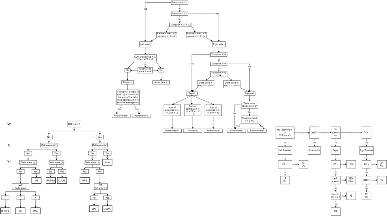
Is there a need for a novel algorithm for accessory pathways localization?Malek Nayfeh MD, Marwan M. Refaat MDDivision of Cardiology, Department of Internal Medicine, American University of Beirut Medical Center, Beirut, LebanonRunning Title: Is there a need for a novel algorithm for WPW localization?Words: 572 (excluding the title page and references)Keywords: accessory pathways, Wolff-Parkinson-white, WPW, cardiac arrhythmias, cardiovascular diseases, heart diseases, Inferior Lead DiscordanceFunding: NoneDisclosures: NoneCorresponding Author:Marwan M. Refaat, MD, FACC, FAHA, FHRS, FASE, FESC, FACP, FRCPAssociate Professor of MedicineDirector, Cardiovascular Fellowship ProgramDepartment of Internal Medicine, Cardiovascular Medicine/Cardiac ElectrophysiologyDepartment of Biochemistry and Molecular GeneticsAmerican University of Beirut Faculty of Medicine and Medical CenterPO Box 11-0236, Riad El-Solh 1107 2020- Beirut, LebanonUS Address: 3 Dag Hammarskjold Plaza, 8th Floor, New York, NY 10017, USAOffice: +961-1-350000/+961-1-374374 Extension 5353 or Extension 5366 (Direct)Wolff Parkinson White Syndrome (WPW) affects between 0.1% and 0.2% of the population, causes morbidity due to supraventricular tachycardia (SVT) and can lead to sudden cardiac arrest [1-3]. The management involves localizing the accessory pathway, and then ablating it, by using either radiofrequency (RF) ablation or cryoablation. The electrocardiogram has been useful over the last decades in the localization of accessory pathways, premature ventricular contractions site of origin and pacing sites [4]. Regarding localization of the pathway, following a large study of RF ablation, Fitzpatrick et al described eight anatomical locations of different pathways using fluoroscopic landmarks: Right anteroseptal (RAS), right midseptal (RMS), right posteroseptal (RPS), right anterolateral (RAL), right posterolateral (RPL), left anterolateral (LAL), left posterolateral (LPL) and left posteroseptal (LPS) [5]. Other algorithms such as the Arruda algorithm or the D’Avila algorithm are also used by clinicians [6, 7]. Most of the accessory pathways’ localization algorithms involve assessment of the delta wave vector (Figure 1), some focus more on QRS morphology (Figure 2), and others combine both methods (Figure 3) [8-12]. By using these algorithms, differentiating between right sided and left sided accessory pathways does not generally pose a problem. However, determining the exact location of right and left sided pathways appears to be more challenging.The study of Bera et al. is a retrospective cohort. Twenty-two patients met the inclusion criteria. The aim was to assess the value of inferior lead discordance (meaning a positive QRS in lead II and a negative QRS in lead III) as a predictor of right anterior (RA) and RAL pathway. The authors included participants who had undergone RF ablation and were found to have right sided pathways. They then separated them in two groups based on if they had RA and RAL pathway (group 1) vs other pathways (group 2). The study found that all patients who had RA and RAL pathway had an ECG showing ILD, while 17 out of 18 patients who were in the other locations did not have an ECG with ILD. The sensitivity and specificity of ILD for predicting RAL location are 100% and 95% respectively.The findings in this study are highly relevant because they represent a clear and simple way of localizing RA/RAL pathways. Other algorithms are also extremely helpful but have their limitations especially if they rely on the delta wave polarity and the electrocardiogram is not fully pre-excited. Another advantage to the algorithm used in this study is that it focuses on limb leads, instead of pericardial leads, which are highly susceptible to variability due to possible displacement.This was a well conducted study, but has some limitations, most notably the small sample size of 22, with only 4 being RA and RAL pathways. There are many algorithms that help cardiologists and cardiac electrophysiologists in localizing accessory pathways before ablation, however, none has specifically focused on RA and RAL pathways. With the advances in artificial intelligence and machine learning, more algorithms using them might be developed in the future.Figure LegendsFigure 1: Examples of algorithms that rely on delta wave polarity such as Fitzpatrick (top) [5], Chiang (bottom left) [8] and Arruda (bottom right) [6]Figure 2: Example of algorithms that rely on QRS morphology such as D’Avilla (top left) [7], Taguchi (top right) [9] and St George’s (bottom) [10].Figure 3: Examples of algorithms that rely both on delta waves and QRS morphology, such as Pambrun (top) [11] and Baek (bottom) [12].References:1. Refaat MM, Hotait M, Tseng ZH (2014). Utility of the Exercise Electrocardiogram Testing in Sudden Cardiac Death Risk Stratification. Ann Noninvasive Electrocardiol, 19(4): 311-318.2. Lu, C. W., Wu, M. H., Chen, H. C., Kao, F. Y., & Huang, S. K. (2014). Epidemiological profile of Wolff-Parkinson-White syndrome in a general population younger than 50 years of age in an era of radiofrequency catheter ablation. International journal of cardiology, 174(3), 530–534. https://doi.org/10.1016/j.ijcard.2014.04.1343. Arai, A., & Kron, J. (1990). Current management of the Wolff-Parkinson-White syndrome. The Western journal of medicine, 152(4), 383–391.4. Refaat M, Mansour M, Singh JP, Ruskin JN, Heist EK (2011). Electrocardiographic Characteristics in Right Ventricular Versus Biventricular Pacing in Patients With Paced Right Bundle Branch Block QRS Pattern. J Electrocardiol, 44 (2): 289-95.5. Fitzpatrick, A. P., Gonzales, R. P., Lesh, M. D., Modin, G. W., Lee, R. J., & Scheinman, M. M. (1994). New algorithm for the localization of accessory atrioventricular connections using a baseline electrocardiogram. Journal of the American College of Cardiology, 23(1), 107–116. https://doi.org/10.1016/0735-1097(94)90508-86. Arruda, M. S., McClelland, J. H., Wang, X., Beckman, K. J., Widman, L. E., Gonzalez, M. D., Nakagawa, H., Lazzara, R., & Jackman, W. M. (1998). Development and validation of an ECG algorithm for identifying accessory pathway ablation site in Wolff-Parkinson-White syndrome. Journal of cardiovascular electrophysiology, 9(1), 2–12. https://doi.org/10.1111/j.1540-8167.1998.tb00861.x7. d’Avila, A., Brugada, J., Skeberis, V., Andries, E., Sosa, E., & Brugada, P. (1995). A fast and reliable algorithm to localize accessory pathways based on the polarity of the QRS complex on the surface ECG during sinus rhythm. Pacing and clinical electrophysiology : PACE, 18(9 Pt 1), 1615–1627. https://doi.org/10.1111/j.1540-8159.1995.tb06983.x8. Chiang, C. E., Chen, S. A., Teo, W. S., Tsai, D. S., Wu, T. J., Cheng, C. C., Chiou, C. W., Tai, C. T., Lee, S. H., & Chen, C. Y. (1995). An accurate stepwise electrocardiographic algorithm for localization of accessory pathways in patients with Wolff-Parkinson-White syndrome from a comprehensive analysis of delta waves and R/S ratio during sinus rhythm. The American journal of cardiology, 76(1), 40–46. https://doi.org/10.1016/s0002-9149(99)80798-x9. Taguchi, N., Yoshida, N., Inden, Y., Yamamoto, T., Miyata, S., Fujita, M., Yokoi, K., Kyo, S., Shimano, M., Hirai, M., &amp; Murohara, T. (2013, December 22). A simple algorithm for localizing accessory pathways in patients with Wolff-Parkinson-White syndrome using only the R/S ratio. Journal of Arrhythmia. Retrieved February 10, 2022, from https://www.sciencedirect.com/science/article/pii/S188042761300165810. Xie, B., Heald, S. C., Bashir, Y., Katritsis, D., Murgatroyd, F. D., Camm, A. J., Rowland, E., & Ward, D. E. (1994). Localization of accessory pathways from the 12-lead electrocardiogram using a new algorithm. The American journal of cardiology, 74(2), 161–165. https://doi.org/10.1016/0002-9149(94)90090-611. Pambrun, T., El Bouazzaoui, R., Combes, N., Combes, S., Sousa, P., Le Bloa, M., Massoullié, G., Cheniti, G., Martin, R., Pillois, X., Duchateau, J., Sacher, F., Hocini, M., Jaïs, P., Derval, N., Bortone, A., Boveda, S., Denis, A., Haïssaguerre, M., & Albenque, J. P. (2018). Maximal Pre-Excitation Based Algorithm for Localization of Manifest Accessory Pathways in Adults. JACC. Clinical electrophysiology, 4(8), 1052–1061. https://doi.org/10.1016/j.jacep.2018.03.01812. Baek, S. M., Song, M. K., Uhm, J. S., Kim, G. B., & Bae, E. J. (2020). New algorithm for accessory pathway localization focused on screening septal pathways in pediatric patients with Wolff-Parkinson-White syndrome. Heart rhythm, 17(12), 2172–2179. https://doi.org/10.1016/j.hrthm.2020.07.016.

Andrew Hughey

and 15 more

Introduction The reuse of cardiac implantable electronic devices may help increase access to these therapies in low- and middle-income countries (LMICs). No published data exist regarding the views of patients and family members in LMICs regarding this practice. Methods and Results A paper questionnaire eliciting attitudes regarding pacemaker reuse was administered to ambulatory adult patients and patients’ family members at outpatient clinics at Centro Nacional Cardiologia in Managua, Nicaragua, Indus Hospital in Karachi, Pakistan, Hospital Carlos Andrade Marín and Hospital Eugenio Espejo in Quito, Ecuador, and American University of Beirut Medical Center in Beirut, Lebanon. There were 945 responses (Nicaragua – 100; Pakistan – 493; Ecuador – 252; Lebanon – 100). A majority of respondents agreed or strongly agreed that they would be willing to accept a reused pacemaker if risks were similar to a new device (707, 75%), if there were a higher risk of device failure compared to a new device (584, 70%), or if there were a higher risk of infection compared to a new device (458, 56%). A large majority would be willing to donate their own pacemaker at the time of their death (884, 96%) or the device of a family member (805, 93%). Respondents who were unable to afford a new device were more likely to be willing to accept a reused device (79% vs. 63%, P<0.001). Conclusions Patients and their family members support the concept of pacemaker reuse for patients who cannot afford new devices.